
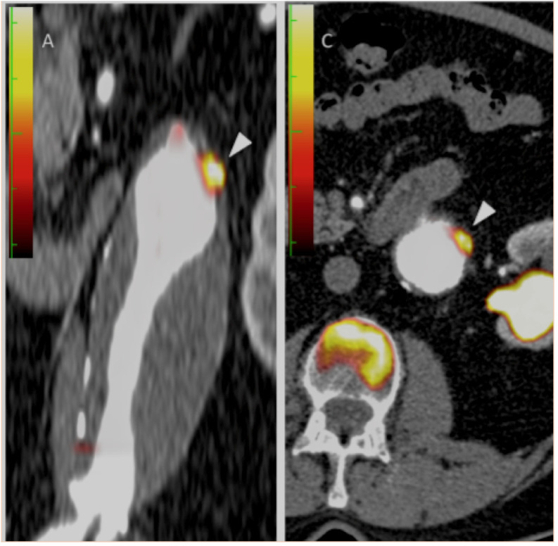
Figure 1.
Coronal and transaxial fused positron emission tomography and contrast CT images demonstrating focal 18F-FDG uptake within the aneurysmal wall (white arrowheads). Note also the calcified lateral aspect of the aneurysm.
Official websites use .gov
A
.gov website belongs to an official
government organization in the United States.
Secure .gov websites use HTTPS
A lock (
) or https:// means you've safely
connected to the .gov website. Share sensitive
information only on official, secure websites.
Coronal and transaxial fused positron emission tomography and contrast CT images demonstrating focal 18F-FDG uptake within the aneurysmal wall (white arrowheads). Note also the calcified lateral aspect of the aneurysm.