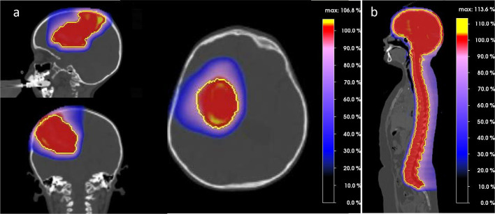
Figure 1.
Sagittal, axial and coronal views of proton dose distribution for a parieto-frontal tumor. PTV is shown in yellow. Figure 1b. Sagittal view of proton dose distribution for craniospinal irradiation. PTV is shown in yellow.Noteworthy, the color wash dose level display all dose levels. As such, absence of colors equals to absence of dose.

