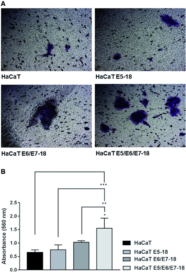
Fig. 3: effect of human papillomavirus (HPV)-18 E5, E6, and E7 oncogenes on invasion in HaCaT cells. Invasive potential was assessed using a QCM™ Collagen Cell Invasion Assay. (A) A total of 9.0 × 10⁵ starved cells were seeded into each chamber containing serum-free dulbecco’s modified eagle’s medium (DMEM) low glucose medium. The bottom chamber was filled with DMEM low glucose 10% foetal bovine serum (FBS) that acted as a chemoattractant, and the plates were incubated at 37°C under 5% CO2 for 48 h. Invading cells were visualised under a Leitz Labovert FS inverted microscope (Oberkochen, Germany) at a magnification of 10x. Two independent experiments were performed in triplicate, and the magnification was 10x (B). Quantification of results obtained in (A) measured by absorbance at 560 nm. The mean ± standard deviation (SD) is shown. A one-way analysis of variance (ANOVA) test was conducted to assess statistical significance among groups along with Tukey’s HSD test for intra-group comparison. (*) p < 0.05, (**) p < 0.01, (***) p < 0.001.

