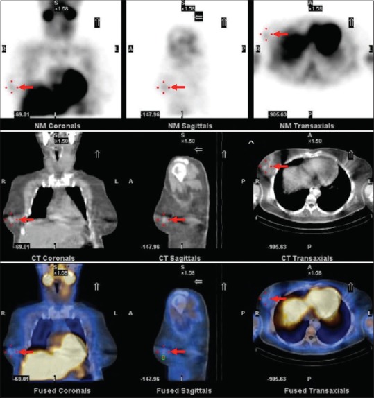
Figure 1.

The planar images of technetium-99m methoxyisobutylisonitrile of the neck and chest (upper row) showed an incidental finding of focal uptake at right breast (arrow). Noncontrast computed tomography of the chest (middle row) demonstrated a 1.3-cm breast nodule at right breast corresponding with the technetium-99m methoxyisobutylisonitrile lesion. Further inspection of the corresponding fusion images of single-photon emission computed tomography and computed tomography (lower row) showed nonfunctioning breast nodule
