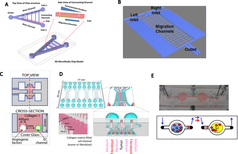
Figure.2.
Tumor-on-a-chip models to induce chemotaxis and study cell-cell crosstalk. A. A V-shape double-channel microfluidic device for chemotaxis. Inlet 1 was designed for media flow and inlet 2 for chemokine perfusion. (reproduced with permission from ref. 32) B. Microfluidic channel design for single-cell chemotaxis. Chemical gradients were generated from the right channel to the left channel. (reproduced with permission from ref. 36) C. Parallel-channels in a 3D collagen scaffold to induce a response of endothelial cells in an angiogenic factors gradient. (reproduced with permission from ref. 39) D. Tumor spheroids and CAFs co-cultured in a 3D hydrogel-based microfluidic chip, separated with medium channels. (reproduced with permission from ref. 46) E. A metastasis-on-a-chip model with a gut organoid and a liver organoid located in two connected PDMS chambers. (reproduced with permission from ref. 50)

