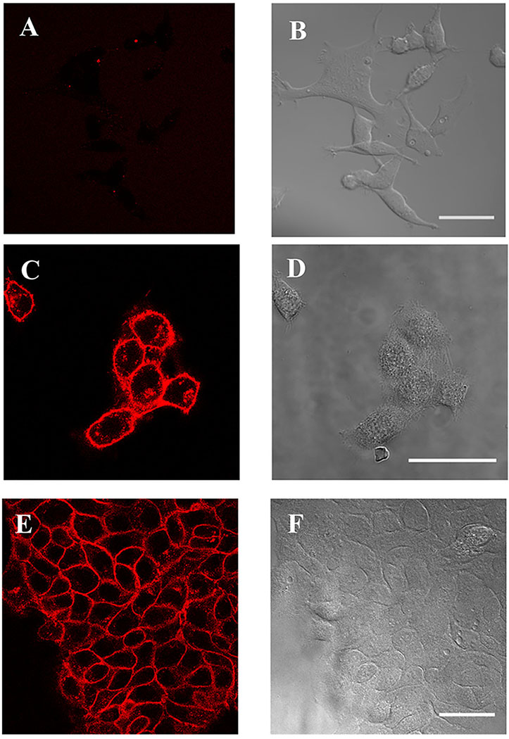
Figure 1.

dL5**-cetuximab labels EGFR on HaCaT and A431 cells. (A-B) HEK-293 cells are not labeled by the reagent. (C-D) HaCaT and (E-F) A431 cells are labeled with dL5**-cetuximab and MG fluorogen. MG fluorescence is shown on the left, DIC transmitted light images on the right. Scale bars = 50μm.
