
Figure 2.
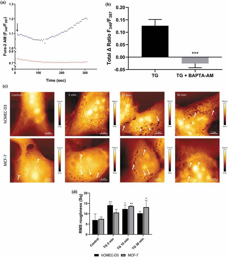
Increasing intracellular calcium with thapsigargin (TG) induces plasma membrane EV biogenesis in malignant MCF-7 cells.
MCF-7 cells were treated with TG (300 nM) for 5 min in the presence and absence of BAPTA-AM (50 uM). Changes to intracellular calcium was analysed using the ImageXpress high content imaging system and ratiometric calcium indicator Fura-2 AM. (a) Representative trace of change in relative fluorescence of Fura-2 AM following treatment with TG alone (●) and in the presence of BAPTA-AM (▴) in MCF-7 cells. The rise in fluorescence indicates increasing intracellular Ca2+ over 5 min. (b) Total TG-mediated increase in intracellular Ca2+ ± BAPTA-AM indicated by increase in relative fluorescence of Fura-2 AM (340/387) in MCF-7 cells. Data represents the mean ± SD of three experiments. ***p < 0.001 (paired t test). (c) Time course of TG-mediated increase in plasma membrane EV biogenesis. hCMEC-D3 (upper panel) and MCF-7 (lower panel) cells treated for 5, 10, and 30 min with TG and EV-derived pits visualized with AFM (arrows). (d) Analysis of hCMEC-D3 and MCF-7 cell topography illustrates there is an increase in surface roughness following thapsigargin treatment. Representative images shown from at least 3 experiments. Data represent the mean ± SD of at least 3 experiments. *p < 0.05, **p < 0.005 (one way ANOVA).