
Figure 4.
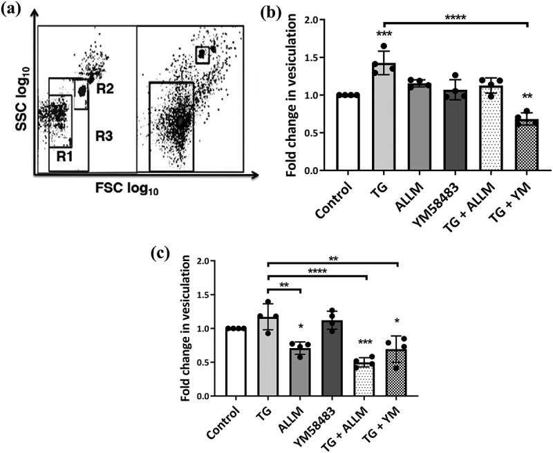
Vesiculation in malignant and non-malignant cells following manipulation of the calcium-calpain plasma membrane EV biogenic pathway.
hCMEC-D3 and MCF-7 cells were treated with modulators of vesiculation and Ca2+ for 24 h EV release was quantified by flow cytometry. (a) EV size gating strategy. Latex sizing beads of (0.3–1.1 um diameter) were used to define the gate for EVs. R1 represents the lower (0.3 µm) and R2 represents the upper (1.1 µm) limits set by the beads. R3 defines the region that was used to quantitate EVs and was applied to all samples. The total number of acquired events when the stop gate was set at 5000 TruCount bead counts is shown on the right. Data are expressed as fold change of EV release relative to vehicle control. (b) Non-malignant hCMEC-D3 cells displayed a significant increase in vesiculation following thapsigargin (TG) treatment and a significant reduction in vesiculation when treated with TG and YM58483. While treatments produced a consistent, small increase in vesiculation, it did not reach statistical significance. (c) Treatment with TG only resulted in a modest increase in vesiculation in malignant MCF-7 cells compared to vehicle control. There were significant reductions in malignant cell vesiculation following treatment with calpain inhibitor II (ALLM), TG + ALLM and TG + YM58483. Data represents the mean ± SD of at least 3 experiments. *p < 0.05, **p < 0.005 ***p < 0.001 (one way ANOVA).