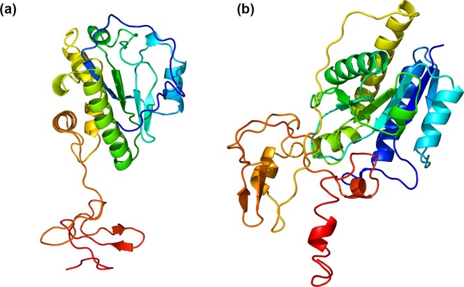
Fig. 5.
Predicted protein structure of the N-acetylmuramoyl-l-alanine amidase of phage Xenia (a) and phage Halcyone (b). The structures were obtained with Phyre2 using the ‘Intensive’ setting. The protein structures are coloured from the C-terminus (red) to the N-terminus (blue). The distinct C-terminal cell-wall binding domain and N-terminal catalytic domain are clearly distinguishable along with the linker connecting them. The amidase of phage Halcyone has a more complex structure.

