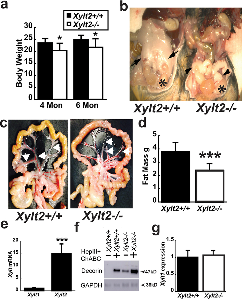
Figure 1.
Body weight, adipose tissue levels, and XylT2 expression in Xylt2−/− mice. (a) Female Xylt2−/− mice on normal chow diet have reduced body weight at 4 and 6 months of age (n=5–8 of each genotype) and (b) gonadal adipose tissue is reduced in 3 month old male Xylt2−/− mice. Arrows in Xylt2+/+ are gonadal adipose tissue and arrowheads in Xylt2−/− show remnants of gonadal adipose tissue. Asterisks indicate testicles. (c) Perivascular mesenteric adipose tissue also reduced in 4-month-old Xylt2−/− females. (d) DEXA scanning analyses shows reduced total body fat in female 4–5 month Xylt2−/−mice, (n=10 of each genotype). (e) Xylt2 is predominant expressed isoform in white adipose gonadal tissue by real time RT-PCR. (f) GAT western from both mice showing free decorin in Xylt2−/− mice. (g) Xylt1 expression did not increase in white gonadal adipose tissue in Xylt2−/− mice, n= 3–4, 2–3 months of age. All analyses are student’s t test where * p<0.05, ***p<0.001.

