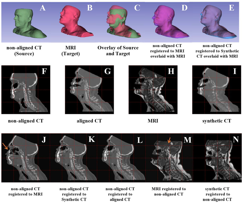
Figure 2.
An example patient shows the various registrations studied in this paper. First row: registration of non-aligned CT (green) to an MR image (red). Box D shows the directly registered non-aligned CT (purple); Box E shows registration of the corersponding synthetic CT to the nonaligned CT. Second row: sagittal view. Boxes F-I are the non-deformed volumes. The third row shows the results for various registrations. Note that the images used in the registration were downsampled to match the 256×256 resolution output from the neural network. All slices shown were in the same location. Arrows denote unanatomical deformation in direct multimodel registration.

