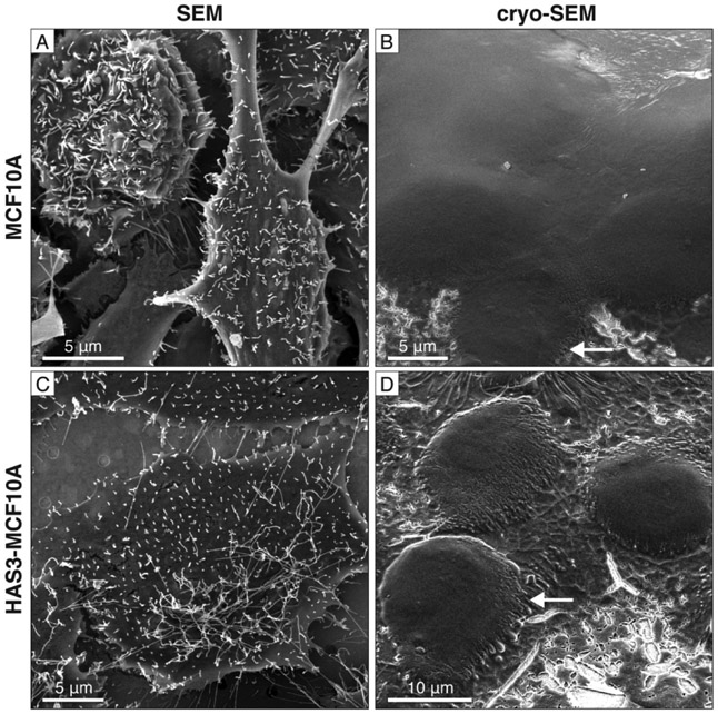
Fig. 2. Cell morphology of HAS3 and MCF10A control cells imaged by conventional and cryo-SEM.
(A) Conventional SEM image of chemically-fixed MCF10A cells. (B) Cryo-SEM image of frozen-hydrated MCF10A cells. Tubular extensions are observed (arrow). (C) HAS3 cells prepared for conventional SEM with longer tubular extensions compared to the MCF10A controls. (D) HAS3 cells imaged by cryo-SEM also show tubular extensions (arrow), but they appear flattened. See Fig. S1 for additional cryo-SEM images of HAS3 cells.

