Figure 5.
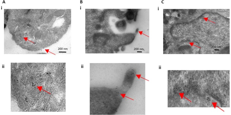
Transmission electron microscopy and immuno-gold labeling of platelet histones. Cells were immuno-gold labeled with anti-histone 4 antibodies and then imaged with transmission electron microscopy. (A,B) Platelets are shown to label positive for histone 4 in their cytoplasm as well as on their cell membrane. Magnified images are shown in the bottom panels (Aii and Bii). (C) HT-29 cells were also immuno-labeled and imaged as a positive control and showed nuclear labeling of histone 4. Magnification of the nuclear envelope with labeling is shown on the bottom panel (Cii). Red arrows point towards immuno-gold particles (6 nm).

