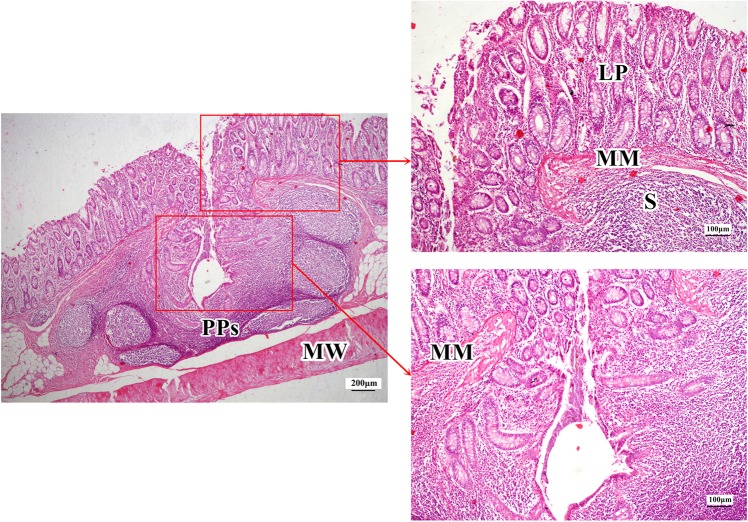
Figure 5.
A crateriform PPs in the midpiece colon of a 42-day old Duan-Nai-An-treated piglet (H&E staining). The inner structure of the PPs appeared scrotiform with well-developed lymphoid tissues on the lateral and basal surfaces of the inner wall of the cyst, well-developed lymphoid follicles near the circular muscle layer, and a large number of scattered lymphocytes in the lamina propria. LP indicates Lamina propria; MM indicates muscularis mucosae; MW indicates muscular wall; PPs indicate Peyer’s Patches; and S indicates Submucosa.

