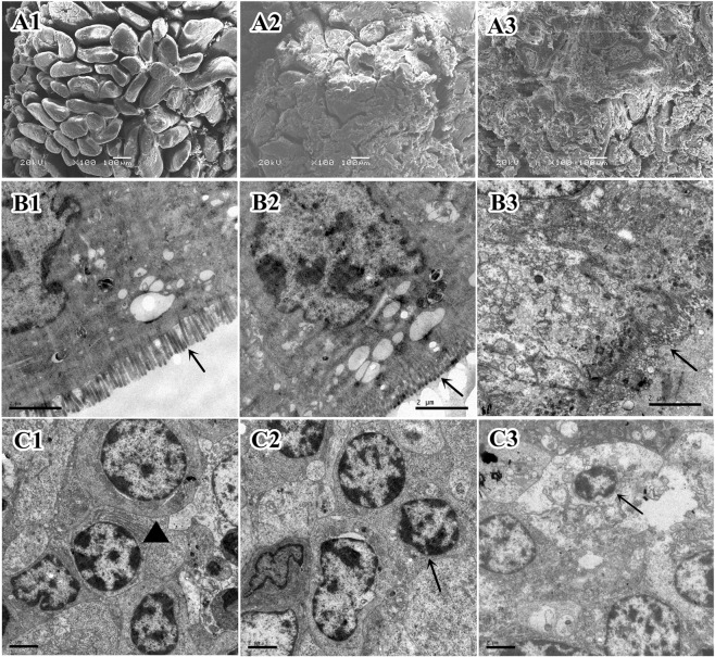
Figure 8.
Electron micrographs of the ileum in 42-day old piglets. (A1) SEM of the ileal mucosa of a Duan-Nai-An-treated piglet showing intact and clearly visible villi. (A2) SEM of the ileal mucosa of a yeast-treated piglet showing localized mucus residues and partial slough of epithelium. (A3) SEM of the ileal mucosa of a control piglet showing a large number of adhering mucus, partial villi damaged, and slough of the epithelium. (B1) TEM of the ileal villi of a Duan-Nai-An -treated piglet showing clear, intact and dense microvilli (indicated by an arrow). (B2) TEM of the ileal villi of a yeast-treated piglet showing intact microvilli (indicated by an arrow). (B3) TEM of the ileal villi of a control piglet showing fractured and sloughed microvilli (indicated by an arrow). (C1) TEM of the ileal lamina propria of a Duan-Nai-An-treated piglet showing well developed lymphocytes with abundant endoplasmic reticulum (indicated by ▲). (C2) TEM of the ileal lamina propria of a yeast-treated piglet showing well developed lymphocytes (indicated by an arrow). (C3) TEM of the ileal lamina propria of a control piglet showing necrosis and chromatin margination of lymphocytes (indicated by an arrow).

