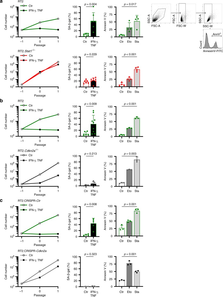
Fig. 3. Stat1- and Cdkn2a-dependent induction of CIS in RT2-cancer cells, but Stat1- and Cdkn2a-independent induction of apoptosis.
a–c Assays were performed with RT2-cancer cells (a, b), RT2.Stat1−/−-cancer cells (a), Cdkn2a-deficient RT2-cancer cells (b), or RT2.CRISPR-Ctr- control, or RT2.CRISPR-Cdkn2a-cancer cells (c). For the senescence growth assay, cells were cultured either with medium (Ctr) or with medium containing 100 ng ml−1 IFN-γ and 10 ng ml−1 TNF for 96 h, washed and then cultured with medium for another 3–4 days. One representative out of 3 independent experiments was given. SA-β-gal activity was determined after 96 h of culture with medium (Ctr) or with medium containing 100 ng ml−1 IFN-γ and 10 ng ml−1 TNF, data show the mean with SD, RT2 Ctr n = 7, CIS n = 7, RT2.Stat1−/− Ctr n = 6, CIS n = 8 (a), RT2 Ctr n = 6, CIS n = 9, RT2.Cdkn2a−/− Ctr n = 4, CIS n = 5 (b), RT2.CRISPR-Ctr Ctr n = 4, CIS n = 5, RT2.CRISPR-Cdkn2a Ctr n = 4, CIS n = 5 (c). For apoptosis induction, cells were exposed to either medium (Ctr) or etoposide (Eto, 100 µM) or staurosporine (Sta, 0.5 µM) for 24 h and then stained for annexin V. Positive cells were detected by flow cytometry data show the mean with SD (a (including gating strategy), b, c), RT2 n = 4, RT2.Stat1−/− n = 4 (a), RT2 Ctr n = 4, Eto, Stau n = 3, RT2.Cdkn2a−/− n = 3 (b), RT2.CRISPR-Ctr and RT2.CRISPR-Cdkn2a n = 3 (c). Significance tested by using unequal variances t-test.

