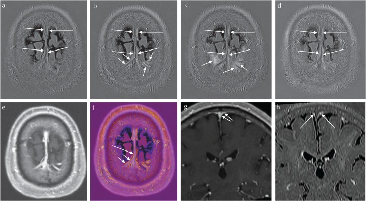
Fig. 2.
A 55-year-old man suspected of endolymphatic hydrops underwent a pre- and post-contrast enhanced MR study. Scan parameters are the same as in Fig. 1. 3D-real IR images were obtained before (a), and at 10 min (b), 4 h (c) and 24 h (d) after intravenous administration of a single dose of gadolinium-based contrast agent (IV-GBCA). The window display level and width were kept constant among (a–d). At 10 min after IV-GBCA, the linear enhancement along the cortical veins and the superior sagittal sinus (SSS) can be seen (arrows and long arrows, b). The linear enhancement along the cortical veins is presumed to be the enhancement in the space between the pial sheath and the wall of the cortical vein (arrows, b). The linear enhancement along the SSS is presumed to be the previously reported meningeal lymphatics (long arrows, a–d). Note the linear enhancement around the cortical vein at 10 min after IV-GBCA (arrows, b) that spreads to the surrounding cerebrospinal fluid (CSF) space at 4 h after IV-GBCA (arrows, c). The enhancement of the meningeal lymphatics along the SSS can be appreciated on images obtained at 10 min after IV-GBCA (b) as well as 4 h after IV-GBCA (c). Magnetization prepared rapid gradient-echo (MP-RAGE) images were obtained at 7 min after IV-GBCA (e). On the MP-RAGE image, the venous lumen is enhanced. The fusion image between (b) and (e) is shown in (f). The enhanced area of the 3D-real IR is colored. On the fusion image, the linear colored enhancement is located along the cortical veins (arrows, f) and the SSS (long arrow, f). The linear colored enhancement is located outside of the venous lumen. The colored linear enhancement along the cortical vein is presumed to be the space between the pial sheath and the cortical venous wall (arrows, f), and appears to continue to the colored enhancement along the SSS (presumed to be the meningeal lymphatics, long arrow, f). This suggests that the space between the pial sheath and the cortical venous wall connects to the meningeal lymphatics. On reformatted coronal MP-RAGE images obtained at 7 min after IV-GBCA (g), a cortical vein drains into the lower part of the SSS (arrows, g). On reformatted coronal 3D-real IR image obtained at 10 min after IV-GBCA, meningeal lymphatics are located at the superior edges of the SSS (arrows, h).

