
Figure 1.
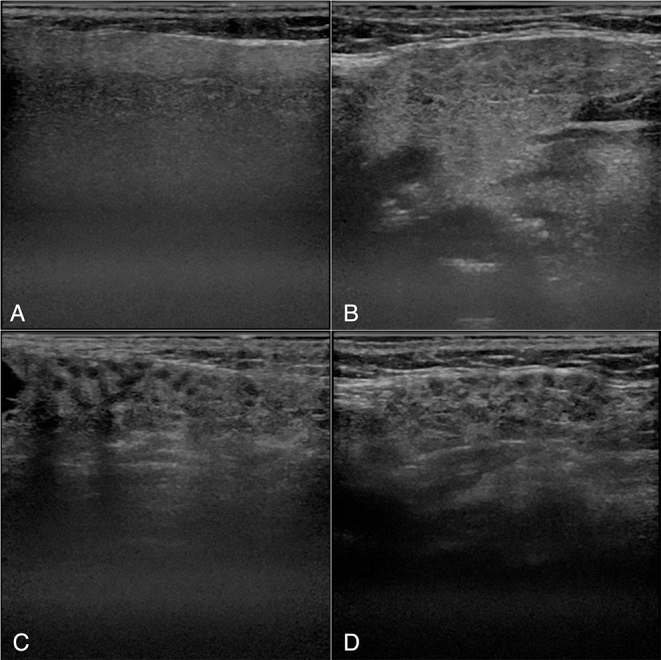
Examples of ultrasonography images. A and B: PG (A) and SMG (B) of a patient with non-SjS. C and D: PG (C) and SMG (D) of a patient with SjS. Inhomogeneous parenchyma characterized by multiple diffuse anechoic regions are observed. SjS, Sjögren’s syndrome.