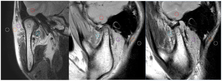
Figure 1. .
Six ROIs from 17 to 20 mm2 in size were drawn on the median oblique sagittal and coronal position, respectively. ROI1, temporal lobe (red circle); ROI2, condylar neck (black); ROI3, lateral pterygoid muscle, LPM (green); ROI4, parotid gland (purple); ROI5, adipose area (orange); and ROI6, the background noise (yellow). LPM,lateral pterygoid muscle; ROI, region of interest.

