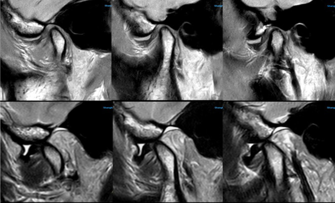
Figure 4. .
It clearly showed the relationship between articular disc and condyle by using Surface coil. TMJ MRI showed anterior disc displacement without reduction and medium amount of effusion in the upper articular cavity, which were obtained by the surface coil (Upper images were OSag PDWI with closed mouth, lower images were OSag T 2WI with opened mouth). The surface coil has better contrast although the signal decays very fast with the increasing in the depth of object, which can make the disc–condyle complex displayed clearly. OSag,oblique sagittal; T 2WI, T 2 weighted image; TMJ, temporomandibular joint.

