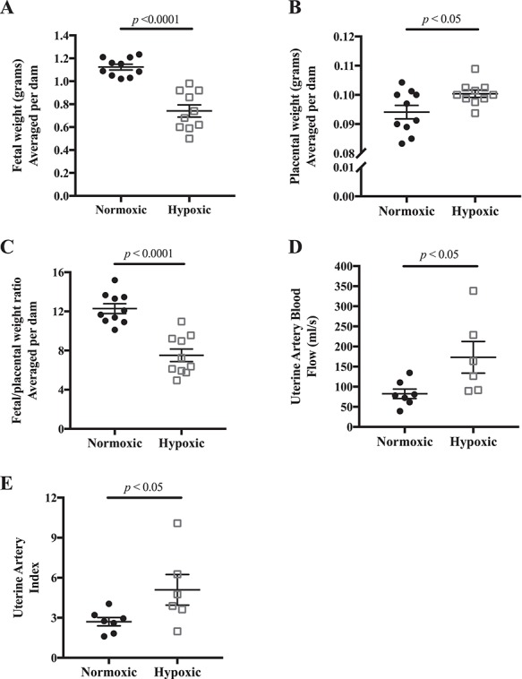
Figure 1.

Effects of late-gestation (E14.5–18.5) hypoxia on fetal growth and uterine artery blood flow. A) E18.5 fetal weight was reduced, B) placental weight was increased, and therefore C) the ratio of fetal to placental weight was lower indicating a selective reduction in fetal growth; n = 10 normoxic, 10 hypoxic dams. D) Uterine artery volumetric blood flow was increased with late-gestation hypoxia, and E) uterine index, the ratio of uterine artery blood flow to cardiac output, was higher; n = 7 normoxic, 6 hypoxic dams. Data are presented as mean ± SEM and analyzed by Student unpaired t-test.
