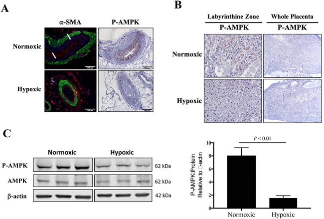
Figure 4.

Characterization of placental and uterine artery AMPK activation. A) By immunohistochemistry, P-AMPK was pronounced in the uterine arterial smooth muscle cells from normoxic mice (n = 3) but not visible in the uterine artery from hypoxic mice (n = 4). In normoxic mice, there was a distinctive layer between the vascular smooth muscle cells (α-SMA, green staining) and endothelium (stained in red with WGA), pointed to by white arrows. B) Immunohistochemistry staining for placental P-AMPK revealed high AMPK activation in the labyrinthine zone of placentas from normoxic mice but was largely absent from hypoxic placentas. C) By western blot, AMPK activation was lower in the total placental homogenate from hypoxic compared to normoxic mice, as compared by Student unpaired t-test (n = 6 each).
