
Figure 1.
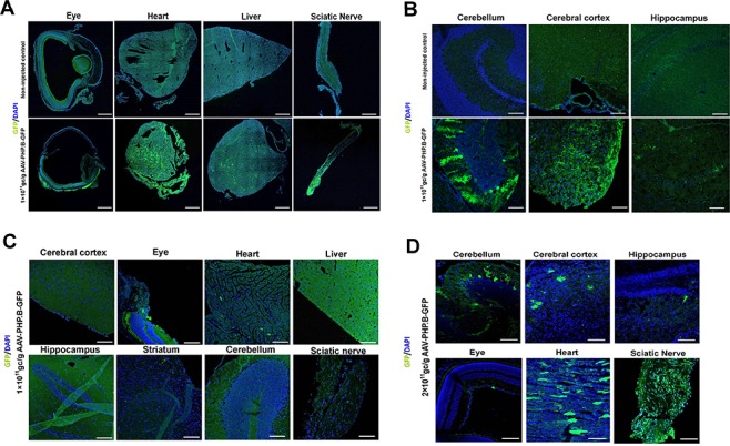
The efficiency of the delivery of AAV–PHP.B-eGFP vector at different dosages and different time points after administration via face vein injection. (A and B) Distribution of the eGFP reporter in peripheral tissues (A) and the central nervous system (B) 2 weeks after face vein injection of the AAV–PHP.B-eGFP vector into neonatal mice. Upper panel, uninjected control; lower panel, mice injected with 1 × 10¹¹ GC/g AAV–PHP.B-eGFP. (C and D) Distribution of eGFP reporter expression in peripheral tissues and the central nervous system 4 weeks after administration of 1 × 10¹¹ GC/g (C) and 2 × 10¹¹ GC/g (D) AAV–PHP.B-eGFP vector via face vein injection.