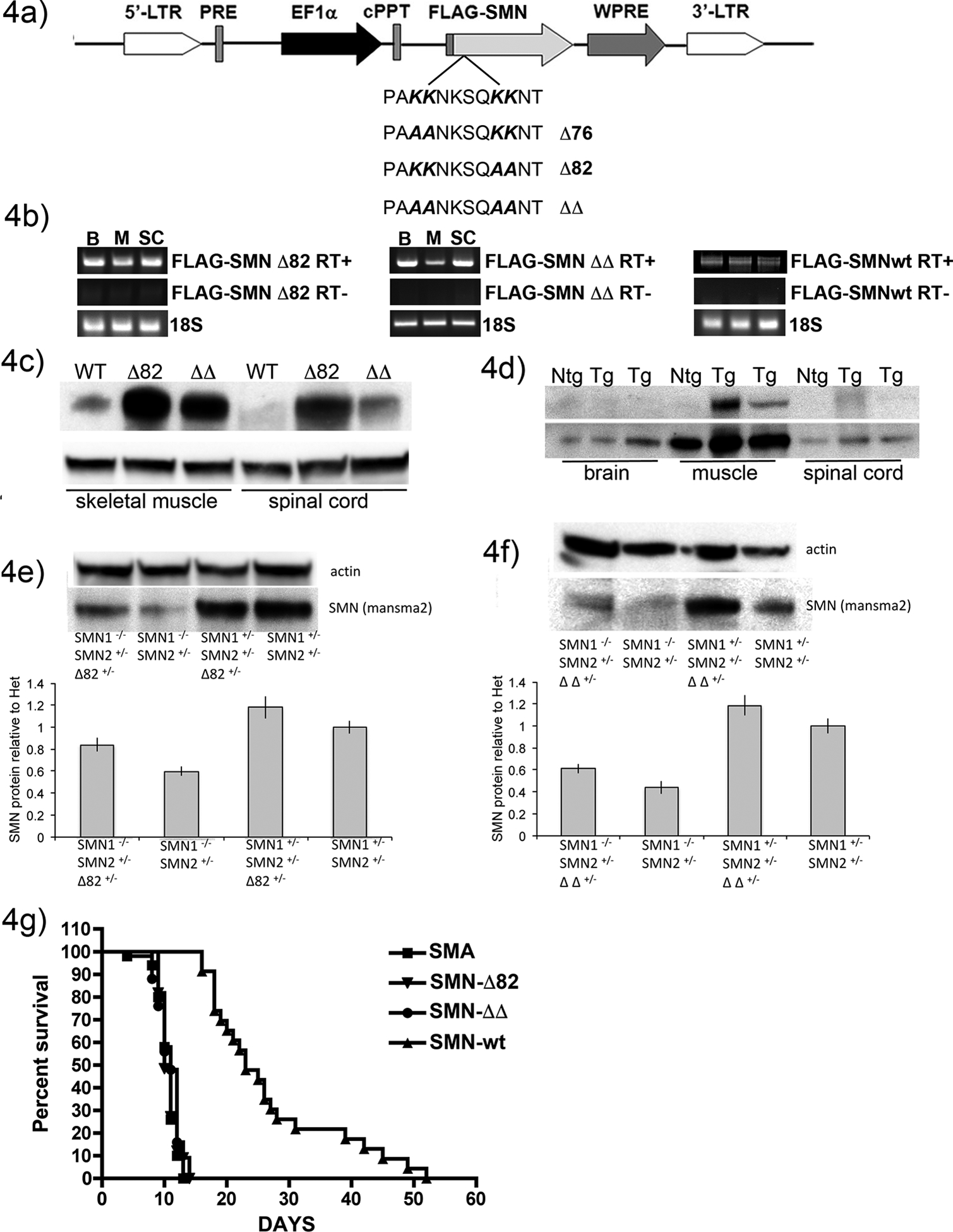
Figure 4: Generation of mice expressing dilysine-mutant SMN.

4a) Schematic of EF1aFLAG-SMN lentiviral vector. 4b) RT-PCR shows transgene expression in brain, muscle and spinal cord from transgene positive F1 progeny in each line. RT- samples were generated with no reverse transcriptase enzyme present demonstrating that no genomic DNA contamination is present. Primers against 18S RNA were used as a loading control to show that equal amount of cDNA were present in each reaction. 4c) Western blot analysis with an antibody specific to human SMN shows FLAG-SMN protein is present in skeletal muscle from all three lines, but only SMN-Δ82 and SMN-ΔΔ protein was detected in spinal cord. 4d) Western blot analysis of brain, muscle and spinal cord lysates from SMN-wt transgenic mice and a non-transgenic sibling confirms that FLAG-SMN protein is only detected in skeletal muscle lysates. 4e) Representative Western blot showing total SMN protein levels in a litter of mice with and without the SMN-Δ82 transgene. SMA pups with the SMN-Δ82 transgene displayed increased levels of SMN although not as high as their healthy siblings. Quantification from multiple experiments (6 animals per genotype) demonstrates a significant increase in SMN protein levels in SMA pups expressing the FLAG-SMN-Δ82 compared to the levels in SMA pups without the transgene (p<0.01 by Student’s t-test). Levels were normalized to non-transgenic healthy littermates using β–actin as a loading control. Error bars represent SEM. 4f) Representative Western blot shows total SMN protein levels in a litter of mice with and without the SMN-ΔΔ transgene. SMA pups with the SMN-ΔΔ transgene show increased levels of SMN, although not as high as their healthy siblings. Quantification from multiple experiments (6 animals per genotype) shows a significant increase in SMN protein levels in SMA pups expressing the FLAG-SMN-ΔΔ compared to the levels in SMA pups without the transgene. (p<0.01 by Student’s t-test) Levels are normalized to non-transgenic healthy littermates using β–actin as a loading control. Error bars represent SEM. 4g) Kaplan-Meier analysis shows that neither SMN-Δ82 nor SMN-ΔΔ transgenes were able to extend survival in the 5058 mouse model. Skeletal muscle expression of FLAG-SMN wt was sufficient to extend survival from 11 to 14 days.
