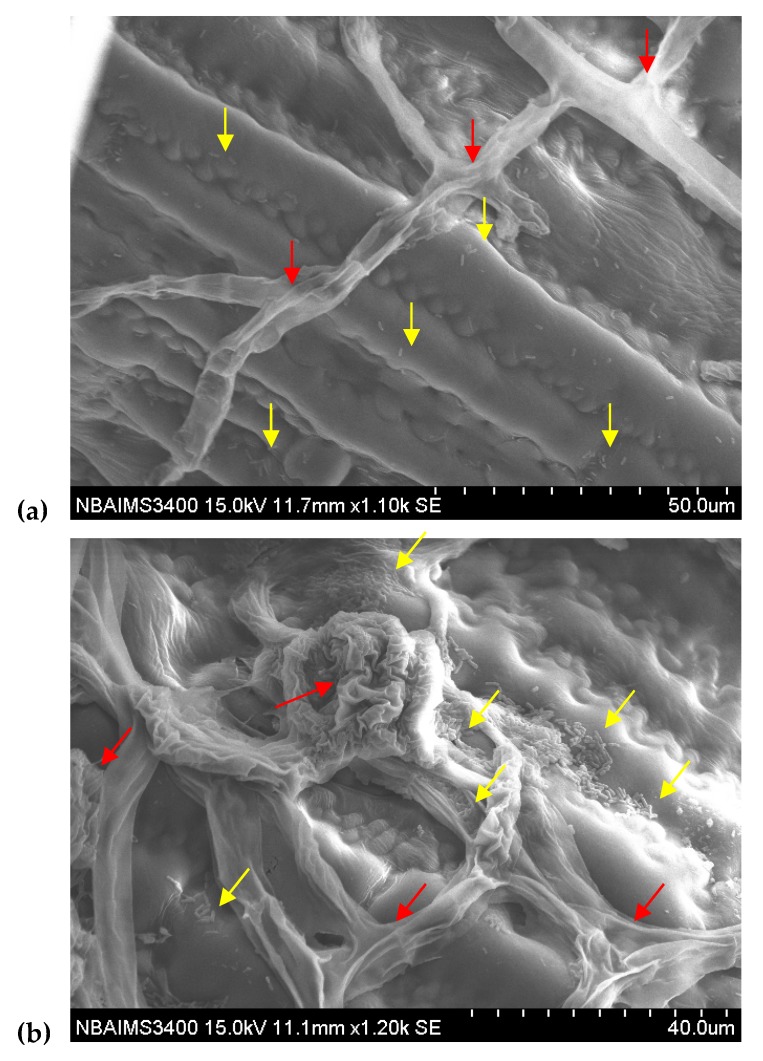
Figure 8.
Scanning electron microscopic study showed the epiphytic colonization of GFP-tagged P. aeruginosa MF-30 on maize leaf. (a) microphotographs taken after 24 h of foliar application, (b) microphotographs taken at 7 days of foliar application. Red arrow indicated the fungal mycelia and infection cushion developed by R. solani on maize leaf, while yellow arrow showed the colonization of P. aeruginosa MF-30 on leaf surface, fungal mycelia, and infection cushion developed on maize leaf. Figure 8 (b) clearly indicates the lysis and disintegration of fungal mycelia and infection cushion caused by MF-30.

