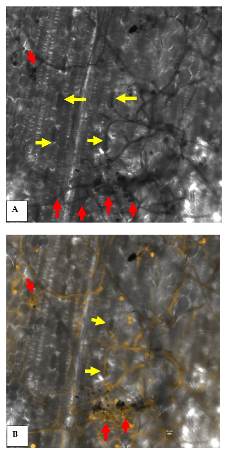
Figure 9.
Confocal scanning laser microphotographs showing the colonization and infection of R. solani in maize. The red arrows indicate the formation of infection cushion, and yellow arrows indicate the entry of fungal mycelia through stomata. (A) Micrograph in TD channel and (B) superimposed image of TD and 488 nm channel together.

