
Figure 2:
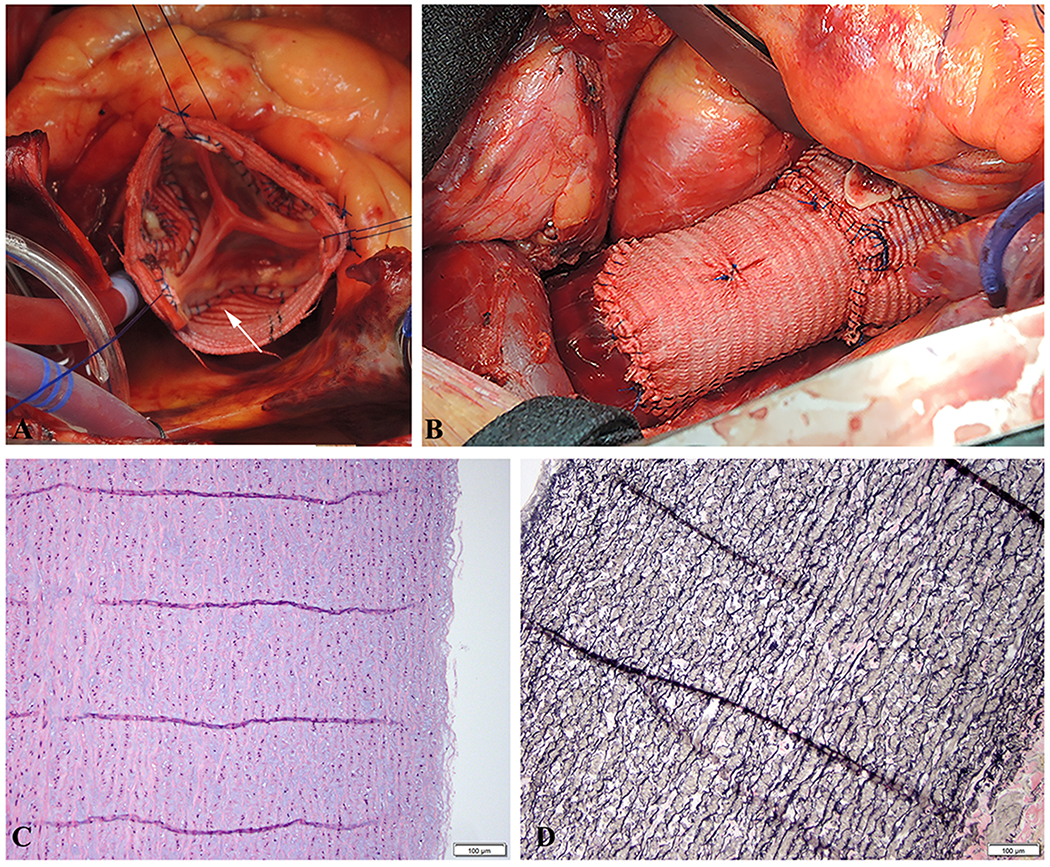
A. Valve-sparing root replacement (David procedure) in a PRKG1 affected patient with a thin and fragile aortic wall. Suture for aortic valve implantation was meticulously placed through the aortic annulus, which was strong enough to hold the suture. The arrow indicates the extremely thin remnant of the aortic sinus wall. B. Completion of the repair showing the distal anastomosis of the Dacron graft to the ascending aorta. C. Moderate, multifocal intralamellar mucoid extracellular matrix accumulation (MEMA-I) without altering the arrangement of the lamellar units. Medial fibrosis and inflammation were also absent. Thus, overall the media displayed mild-to-moderate medial degeneration. Additionally, there was no evidence of aortitis or dissection. (H&E stain). D. The aortic media also displayed moderate, multifocal elastica fragmentation, plus multifocal mild-to-moderate elastic fiber thinning. However, there was no elastic fiber disorganization, no smooth muscle loss or disorganization, and no laminar medial collapse. (EVG stain).