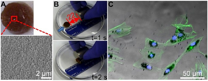
Figure 4.
The characteristics of the magnetic hydrogel. (A) General view of the magnetic hydrogel. The magnified image was obtained by a scanning electron microscope (SEM). (B) Magnetic responses of the magnetic hydrogel. Blue arrows suggested the hydrogel movement orientation before the next time point. (C) Representative images of F-actin (green) by fluorescence microscopy and magnetic nanoparticles (black) by light microscopy showed the endocytosis of MNPs by BMSCs [reproduced with permission from Zhang et al. (2015), Copyright 2015 American Chemical Society].

