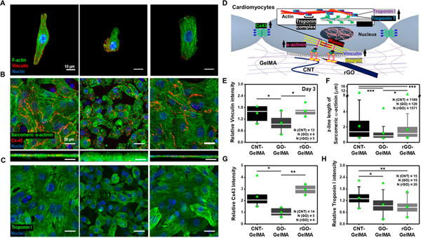
Figure 4.
Phenotypes of cardiac cells cultured on different hybrid scaffolds based on expression of cardiac specific markers. (A) Immunofluorescence images of cardiomyocytes cultured on CNT-, GO-, and rGO-GelMA stained for F-actin (green), vinculin (red), and nuclei (blue). (B) Immunostaining of sarcomeric α-actinin (green), Cx-43 (red), and nuclei (blue) showing cardiac tissues cultured for 5 days on CNT-, GO-, and rGO-GelMA are phenotypically different. Bottom images display cross-section images of cardiac tissues. (C) Immunofluorescence images of cardiomyocytes on CNT-, GO-, and rGO-GelMA stained for troponin I (green) and nuclei (blue). (D) Cartoon showing a schematic representation of part of important proteins for cardiomyocytes during maturation such as integrin (yellow), vinculin (royal), α-actinin (pink), myosin (red), troponin I (purple) and T (blue), and Cx43 (green). (E) Relative intensity of vinculin for cells cultured for 5 days on CNT-, GO-, and rGO-GelMA. (F) Z-line length of sarcomeric α-actinin of cardiomyocytes on different hybrid scaffolds on day 5. Relative intensity of (G) Cx43 and (H) troponin I for cells cultured for 5 days on CNT-, GO-, and rGO-GelMA (*P < 0.05, **P< 0.005, ***P < 0.005). Error bars represent standard deviation.

