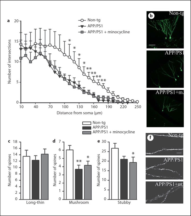
Fig. 4.
Minocycline does not rescue dendritic atrophy and loss of spines in APP/PS1 mice. a Scholl analysis of mature new granular cells labeled with a retroviral vector expressing GFP revealed loss of dendritic complexity in APP/PS1 mice compared to non-tg mice, regardless of the treatment. The graph represents the mean number of intersections between dendrites and concentric radii, centered at the cell body, as a function of distance from the soma, n = 10 cells per group. b Representative confocal pictures of GFP+ mature new neurons for the three groups in study. Computer-assisted classification of spines along 40-µm segments did not detect difference in the number of ‘long-thin’ spines among groups (c); however, the analysis reported lower numbers of ‘mushroom’ spines (d) and ‘stubby’ spines (e) in APP/PS1 mice, both vehicle- and minocycline-treated, compared to non-tg (n = 25 segments per group). f Selected high-magnification segments from respectively non-tg, APP/PS1 vehicle- and minocycline-treated mice. Scale bars = 60 µm (b), 5 µm (f). Error bars represent SEM. * p < 0.05, ** p < 0.001, ANOVA followed by Bonferroni post hoc test.

