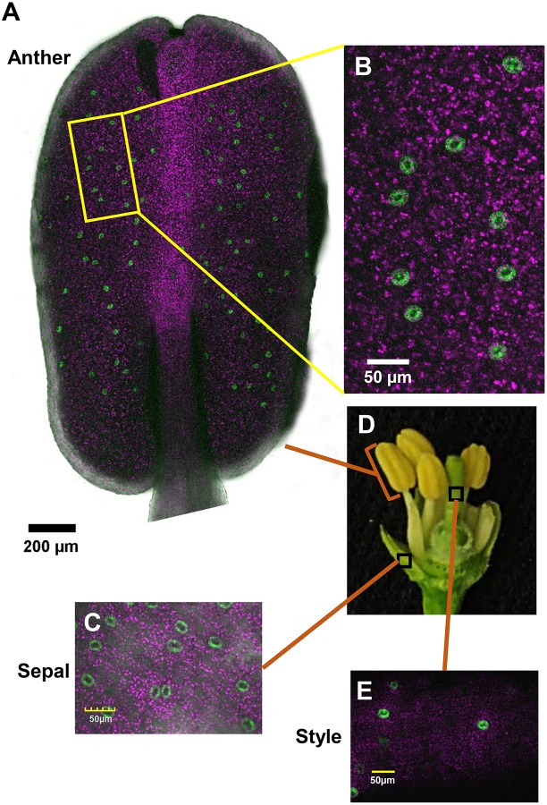
FIGURE 1.
Distribution of stomata on the organs of a closed citrus flower. The confocal images A–C and E are merged images of white light, chlorophyll autofluorescence (stained magenta), and GFP fluorescence (stained green). Flowers were harvested from GCGFP plants. (A) Confocal image of an anther removed from a closed flower. (B) Enlargement of the square in part A. (C) Confocal image of a sepal removed from a closed flower, as indicated in part D. (D) Dissection of a closed flower revealing the floral organs. (E) Confocal image of a style removed from a closed flower. Analyses were performed on five biological replicates.

