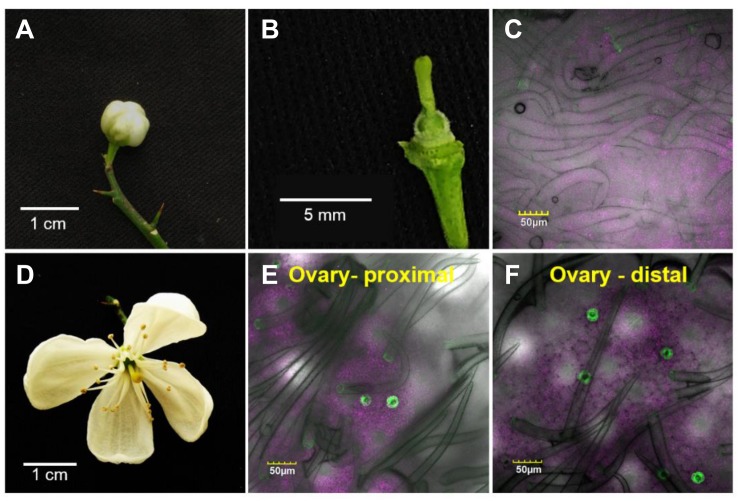
FIGURE 2.
Temporal and spatial distribution of the stomata on a citrus ovary. The confocal images C, E, and F are merged images of white light, chlorophyll autofluorescence (stained magenta), and GFP fluorescence (stained green). Samples were taken from GCGFP plants. The images in the lower row were taken from flowers 3 days older than the images shown in the upper row. (A) A closed GCGFP flower. (B) Binocular image of a dissected closed flower, revealing the ovary. (C) Confocal image of an ovary from a closed flower. (D) An open GCGFP flower. (E) Confocal image of the proximal part of an ovary from an open flower. (F) Confocal image of the distal part of an ovary from an open flower. Analyses were performed on five biological replicates.

