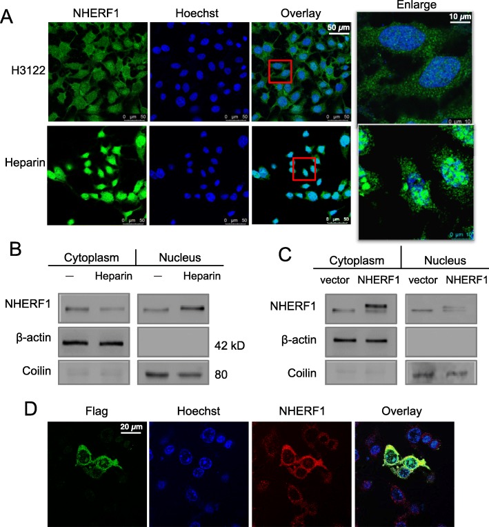
Fig. 7.
Subcellular localization of endogenous, ALK-induced or ectopic overexpressed NHERF1 proteins in H3122 cells. a Immunofluorescent microscopy in native H3122 cells (upper) or cells treated with Heparin (100 μg/ml) for 30 min (lower). b Western blot analyses for cytoplasm or nuclear distribution of NHERF1 proteins, where Coilin was used as a nuclear maker (Full-length blots/gels are presented in Suppl Figure 6). c Western blot of NHERF1 in cells transfected with a Flag-NHERF1 plasmid for ectopic overexpression (Full-length blots/gels are presented in Suppl Figure 6). d Confocal microscopy for distinguishing ectopic NHERF1 vs. the endogenous protein

