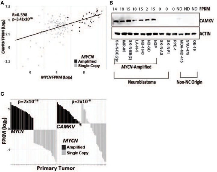
Figure 2.
CAMKV expression correlates with MYCN expression. (A) MYCN and CAMKV FPKM correlate across 150 primary neuroblastoma tumors (R = 0.598, p = 3.41 × 10⁻¹⁶). (B) CAMKV is expressed in MYCN amplified neuroblastoma cell lines (lines 1–7) but not non-amplified lines (8, 9), retinal pigment epithelial cells (9) or non-neural-crest origin cells (11–13). FPKM for CAMKV in cell lines are shown at top, ND = not detected. (C) RNA sequencing data from 150 primary neuroblastoma tumors were stratified by MYCN expression and the FPKM for MYCN (left) and CAMKV (right) for each tumor are plotted. Tumors with amplified MYCN are colored in black and non-amplified tumors are gray. MYCN amplification correlates with MYCN FPKM (p = 2 × 10⁻¹⁶) and CAMKV FPKM (p = 2 × 10⁻⁸).

