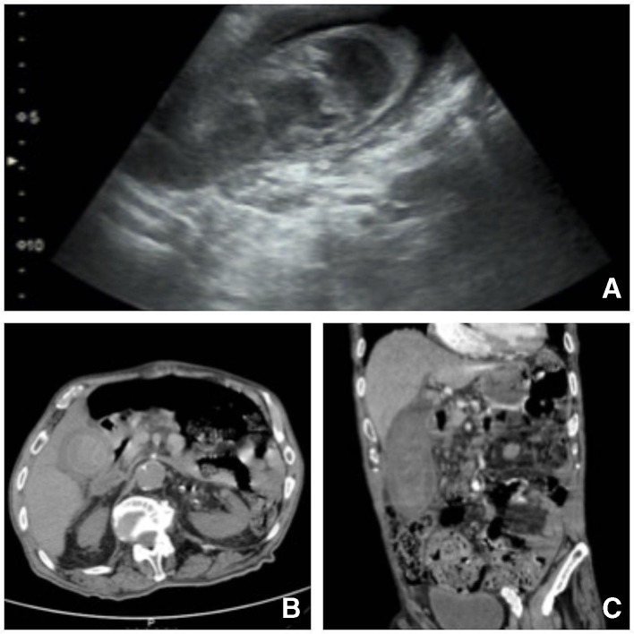
Figure 1.
Ultrasound (A) and CT (B, C) images demonstrating distended gallbladder, thickened/laminated wall, containing bulky image with areas of spontaneous hyperdensity suggesting the presence of a clot in the gallbladder lumen, coexisting perivesicular fat densification, imaging aspects of acute haemorrhagic cholecystitis; there were no signs of active haemorrhage in the lumen of the gallbladder at the time of acquisition of the images; in the infundibulum, an image of spontaneous hyperdensity, which may correspond to the presence of clot versus lithic focus and discrete prominence of the intrahepatic biliary tract.

