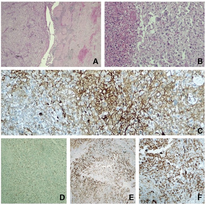
Figure 3.
(A) H&E 40× gallbladder with acute cholecystitis lesions with haemorrhage (right) and where solid pattern neoplasm is identified (left). (B) H&E 200× at higher magnification; the neoplasia consists of epithelioid cells, with marked nuclear pleomorphism and abundant mitosis figures, some atypical, in a dense vascularised stroma with extravasation of erythrocytes. (C) Immunohistochemistry (IHC) Factor VIII. (D) IHC FLI1. (E) IHC AE1AE3. (F) IHC CD31. In our study, focal positivity was observed for Factor VIII, FLI1 and CD31 (vascular markers, which led us to consider it to be an angiossarcoma) and also to cytokeratins AE1AE3 (which is not infrequent in angiosarcoma).

