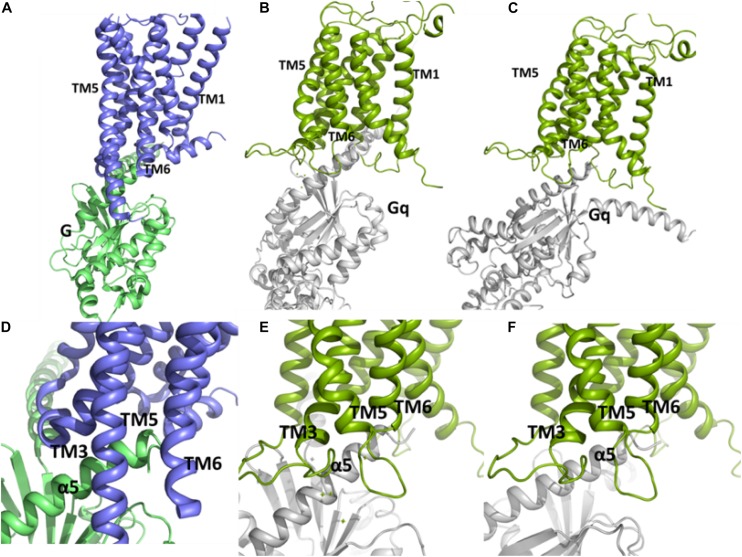
FIGURE 6.
The conformations of the complex between Gq and the TM domain of mGlu5 obtained by using protein-protein docking using Cluspro (B,E) and Haddock (C,F) are similar to the crystallographic β2 adrenergic receptor-G protein complex (PDB ID: 3SN6) (Rasmussen et al., 2011) (A,D). The C terminal of α5 helix within Gα subunit of Gq is located between the intracellular ends of TM3, TM5 and TM6.

