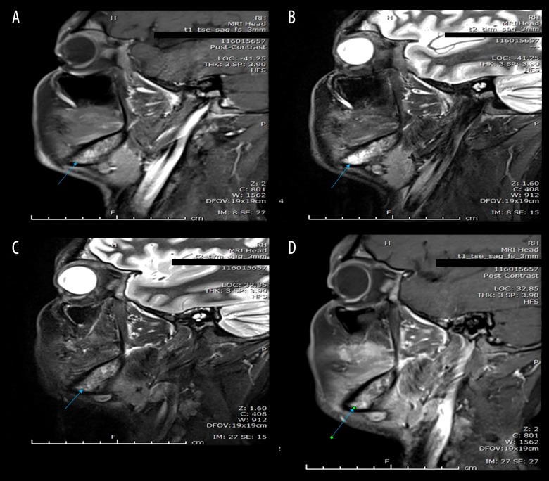
Figure 2.
(A, B) Sagittal STIR and post contrast fat saturated images of right mandible. (C, D) Sagittal STIR and post contrast fat saturated images of left mandible show heterogeneous appearance of bone marrow of mandible with high signal intensity in STIR images and heterogeneous enhancement after intravenous contrast administration (blue arrows) suggesting underlying infiltration.

