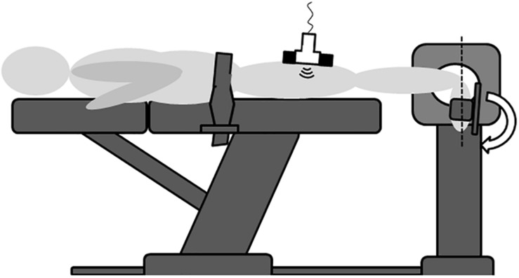
FIGURE 1.
Schematic illustration of the experimental set-up. In the prone position and with the knee extended, the ankle joint was passively moved between plantar flexion and dorsal extension (arrow shows way into dorsal extension). Maximal dorsal extension was calculated as the distance from a calibrated neutral zero position (dashed line). Ultrasound recordings were made over the semimembranosus muscle in order to estimate tissue displacement induced by the ankle motion. EMG (not depicted here) used as biofeedback ensured that no voluntary muscle activity occurred. Pelvic fixation with a strap was required to prevent body motion induced by the dynamometer action. As the belt was not in contact with the Hamstrings, it did not affect tissue displacement.

