
Figure 2.
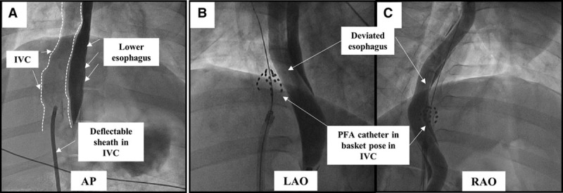
Fluoroscopic view of the esophageal injury mode: pulsed field ablation (PFA) cohort. A, Contrast angiography was performed using a long deflectable sheath placed in the inferior vena cava (IVC; outlined). In the anteroposterior (AP) view, the IVC is seen rightward of the contrast filled esophagus. B and C, Left and right anterior oblique (LAO and RAO) projections demonstrate the PFA catheter in basket pose forcefully pushed against the deviated esophagus. The PFA catheter is shown here ablating 2 different esophageal locations.