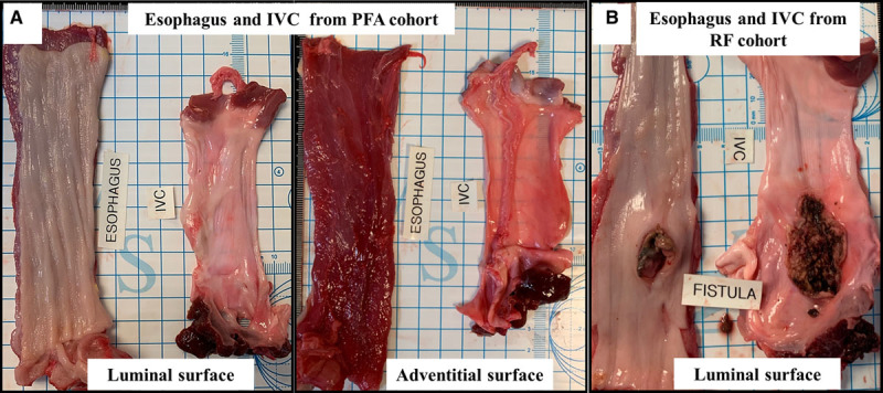
Figure 4.

Gross necropsy views of esophageal changes at sacrifice. A, Representative images demonstrating the normal luminal and adventitial surface of the esophagus after pulsed field ablation (PFA). B, Conversely, from the radiofrequency (RF) ablation cohort, a perforating ulcer is seen on the luminal aspect of the esophagus that was separated from a larger necrotic area opening into the inferior vena cava (IVC; esophagus-IVC fistula).
