
Figure 7.
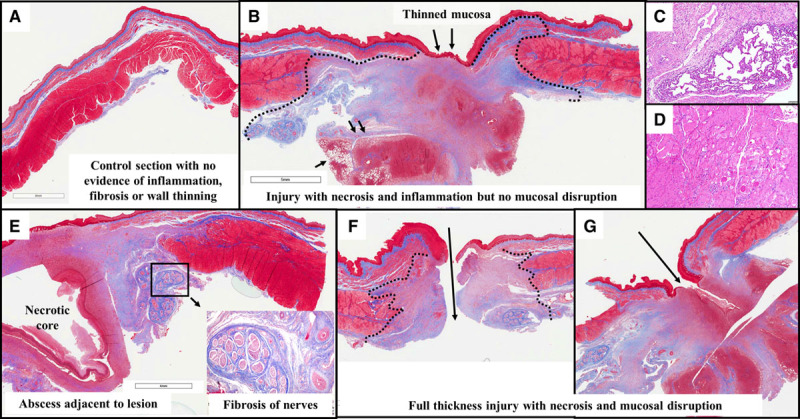
Esophageal histology: radiofrequency ablation (RFA) cohort. A, In this control section from the RFA cohort (ie, from a segment of the esophagus above the level of ablation), there is normal esophageal architecture with no evidence of lesion. B, This section demonstrates near-full-thickness injury with complete necrosis of the muscular layers below the area of mucosal thinning. Granulation tissue is seen extending into the adjacent lung (small black arrows). C, This zoomed (10×) view reveals adherent lung tissue with thickening of alveoli walls and inflammation. D, The tunica muscularis in the vicinity of the lesions shows interstitial inflammation, degenerative vacuolated myocytes, necrotic myocytes (loss of striation, nuclei, eosinophilic cytoplasm), and fibrosis. E, This section reveals a necrotic abscess core within the lesion extending up to the mucosa. The inset focuses on the adventitial lesion resulting in inflammation and fibrosis, involving the periesophageal nerve trunks and thickened arterial walls. F, This section depicts a perforating ulcer with disruption of the wall. G, Here, a fistula tract extends into granulation tissue well beyond the mucosal layer.