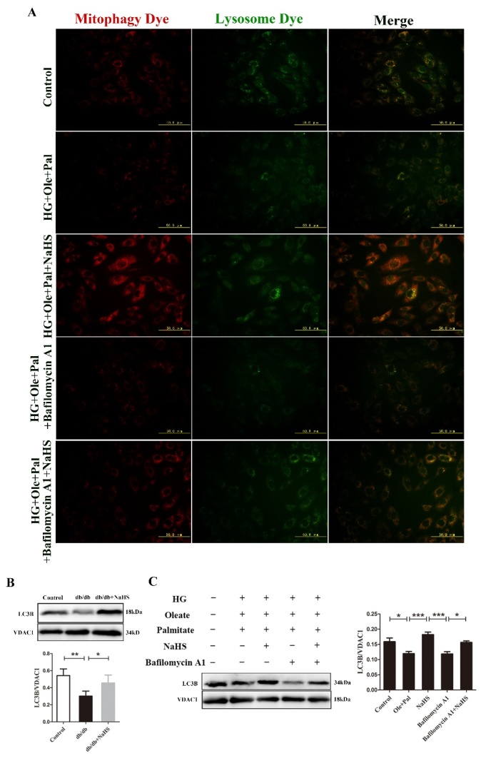
Figure 2.

Exogenous H2S promoted mitophagy in the hearts of db/db mice and in neonatal rat cardiomyocytes. (A) Mitophagosomes were detected in neonatal rat cardiomyocytes by mitophagy detection kit. Red fluorescence represents the mitophagosomes and green fluorescence represents the fusion of mitophagosomes and lysosomes. (B) The expression of LC3B was examined in the mitochondria of db/db cardiac by Western blotting. (C) The expression of LC3B in mitochondria was examined by western blotting following the treatment of neonatal rat cardiomyocytes with Bafilomycin A1. Values are presented as the mean ± S.D. from n = 4 replicates. *P<0.05, **P<0.01, ***P<0.001.
