Figure 2.

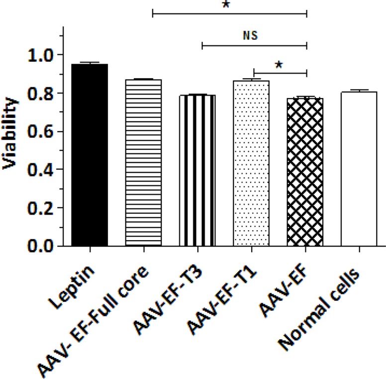
LX-2 cell proliferation assay after transfection with AAV-EF-core full, AAV-EF-T1, and AAV-EF-T3. * P < 0.05 indicated the significant level in comparison with the negative control group (AAV-EF)

LX-2 cell proliferation assay after transfection with AAV-EF-core full, AAV-EF-T1, and AAV-EF-T3. * P < 0.05 indicated the significant level in comparison with the negative control group (AAV-EF)