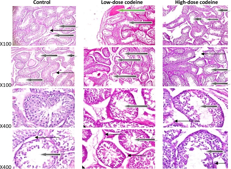
Fig 11. Photomicrographs of testicular sections stained by Haematoxylin and Eosin.
The control shows some normal seminiferous tubules containing normal germ cells including spermatogonia cells and sertoli cells, and normal maturation stages with presence of spermatozoa within their lumen(white arrow). There very few tubules show maturation arrest at secondary stage of maturation (black arrow). The interstitial spaces show normal leydig cells (slender arrow). The rabbits on low-dose codeine show very poor architecture. The seminiferous tubules show double cell layer or thickened propria indicative of cessation of spermatogenesis. They exhibit vacuolation, sloughed germ cells and maturation arrest and show giant cells (black arrow). There is evidence of vascular congestion (green arrow). The interstitial spaces appear normal (slender arrow). The animals on high-dose codeine show very poor architecture. The seminiferous tubules show double cell layer or thickened propria indicative of cessation of spermatogenesis. They exhibit vacuolation, sloughed germ cells and maturation arrest and show giant cells (black arrow). The interstitial spaces appear normal (slender arrow).

