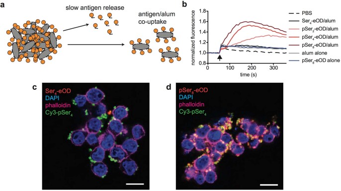
Extended Data Fig. 2. Antigen-specific B cells internalize pSer-antigens bound to alum particles in vitro.
(a) Schematic of potential release of free antigen vs. release of antigen-decorated alum particles at the injection site. (b) glVRC01-expressing human B cells were mixed with eOD (50 nM) and alum (10 µg/mL), or alum alone (10 µg/mL). Shown is calcium signaling in B cells following addition of antigen/alum at 60 sec (arrowhead) by the Fluo-4 fluorescence reporter. (c, d) Alum was labeled by mixing with Cy3-pSer4. glVRC01-expressing B cells were then incubated with fluorescent eOD (50 nM) and fluorophore-tagged alum (10 µg/mL) for 1 hour, and then imaged by confocal microscopy. (scale bars = 10 µm). Experiment was performed in two times, showing representative images from one experiment.

