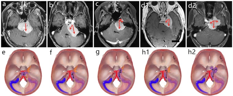
Figure 1.
The proposed classification of PCMs. (a,e) Clivus type: The dural attachment originates from the intradural part of the petroclival fissure, and the main part of the lesion is located in the middle-upper clivus, mainly grows toward the middle line or even the contralateral region. (b,f) Petroclival type: The dural attachment also originates from petroclival fissure, but mainly grows toward the homolateral dorsal petrosum region, and the main part is located in the middle clivus and expands toward the cerebellopontine angle region. (c,g) Petroclivosphenoidal type: The growth pattern is mainly from the posterior cranial fossa to middle cranial fossa; the dural attachment originates from the middle-upper clivus, expands forward and upward along the petroclival fissure, and extends to the dorsum sellae and parasellar region or arrives at the posterior wall of CS. (d1,h1) Sphenopetroclival subtype I: The lesion totally originates within the CS without encroaching the sinus wall resulting in the sinus area expansile hyperplasia; the lateral wall of the lesion is smooth maintaining the dural space between the lesion and temporal lobe. (d2,h2) Sphenopetroclival subtype II: The lesion originates on the lateral wall of CS and grows toward both outside and inside of sinus, and partial lesion could reach the sphenoclival fissure, expand into the parasellar region, and invade into the PCF; the sinus wall is rough and dural space is unclear or even disappeared between the lesion and temporal lobe. The red arrows and black arrows showed the growth pattern of lesions in different types.

