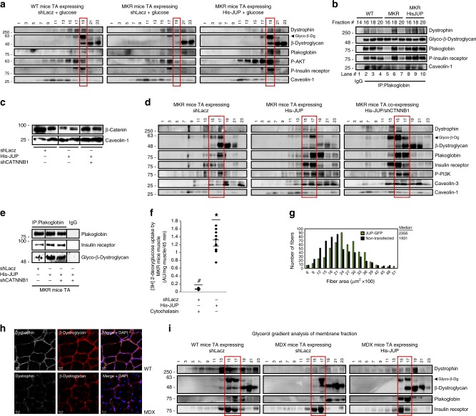
Fig. 5. Overexpression of plakoglobin alone enhances DGC-insulin receptor association and insulin signaling in muscles from MKR or mdx mice.
Muscles from WT or diabetic MKR mice were transfected with 6His-plakoglobin (His-JUP) or control (shLacz) plasmids (contralateral limbs). a Mice were injected with glucose (1 mg/g body weight) and membrane extracts from transfected muscles were analyzed by glycerol gradient and immunoblotting. n = three independent experiments. b Plakoglobin immunoprecipitation from glycerol gradient fractions presented in a. n = two independent experiments. c–e Membrane extracts from muscles expressing shLacz or β-catenin shRNA (shCTNNB1), with or without the co-electroporation of His-plakoglobin, were analyzed by immunoblotting (c), glycerol gradient fractionation (d), and immunoprecipitation (e). Black lines indicate the removal of intervening lanes for clarity and presentation purposes. n = two independent experiments. f [3H]-2-deoxyglucose uptake by muscles expressing shLacz in the presence of cytochalasin B (a competitive inhibitor of regulated glucose transport into cells) or His-JUP is plotted as ratio to shLacz control, which was not treated with cytochalasin B. Data are presented as AU/mg muscle/45 min. Data are mean ± SEM, n = 11 mice for His-JUP and shLacz, n = 4 mice for shLacz treated with cytochalasin B, *P = 0.01 and #P = 0 vs. shLacz in the absence of cytochalasin B, by one-tailed t test. n = two independent experiments. g Overexpression of plakoglobin in muscles from MKR mice attenuates the reduction in fiber size. Measurement of cross-sectional area of 530 fibers expressing His-JUP (also express GFP; green bars) vs. 530 non-transfected fibers (black bars) in the same muscle. Data are acquired from seven mice. h Cross-sections of muscles from WT or mdx mice were immunostained with antibodies against dystrophin and β-dystroglycan (bar, 20 μm). n = two independent experiments from two different mice (representative confocal images are shown). i Membrane extracts from muscles expressing shLacz or His-JUP from WT and mdx mice and analyzed by glycerol gradient fractionation and immunoblotting. Transfected mdx mice muscles are from contralateral limbs. n = two independent experiments.

