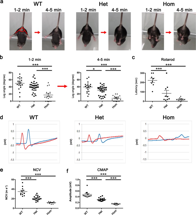
Fig. 1. Motor disturbance and motor nerve dysfunction in L-MPZ mice.
a Representative postures for each mouse genotype at early (1–2 min) and later (4–5 min) time points. To investigate motor function of L-MPZ mice, the tail suspension test was performed for 6 min using 10-week-old L-MPZ wild-type (WT), heterozygous (Het), and homozygous (Hom) mice. Angles formed between bilateral hindlimbs are indicated as red lines in the left image (1–2 min, WT). b Comparison of motor function by bilateral hindlimb angles indicated in a at two time points using video-recorded pictures. c Comparison of motor function examined by the rotarod test. The rotation speed of the rod was 20 rpm over 5 min in each trial. Latency to the time when the mouse fell from the rod was measured. d Representative waveform sets of compound muscle action potentials (CMAPs) obtained from individual genotypes. Motor nerve conduction study was performed in L-MPZ mice. The left or right tibial branch of the sciatic nerve was electrically stimulated at the ankle (d, red line) or sciatic notch (d, blue line). e, f Comparison of average left and right sciatic nerve conduction velocities (NCV; e) and CMAPs (f) obtained from individual genotypes. *p < 0.05; ***p < 0.001 by ANOVA with post-hoc Tukey’s test. Data are presented as mean ± SE of experiments. N in WT = 16, Het = 24, Hom = 14 (b); WT = 8, Het = 9, Hom = 9 (c); and WT = 16, Het = 24, and Hom = 9 (d–f).

