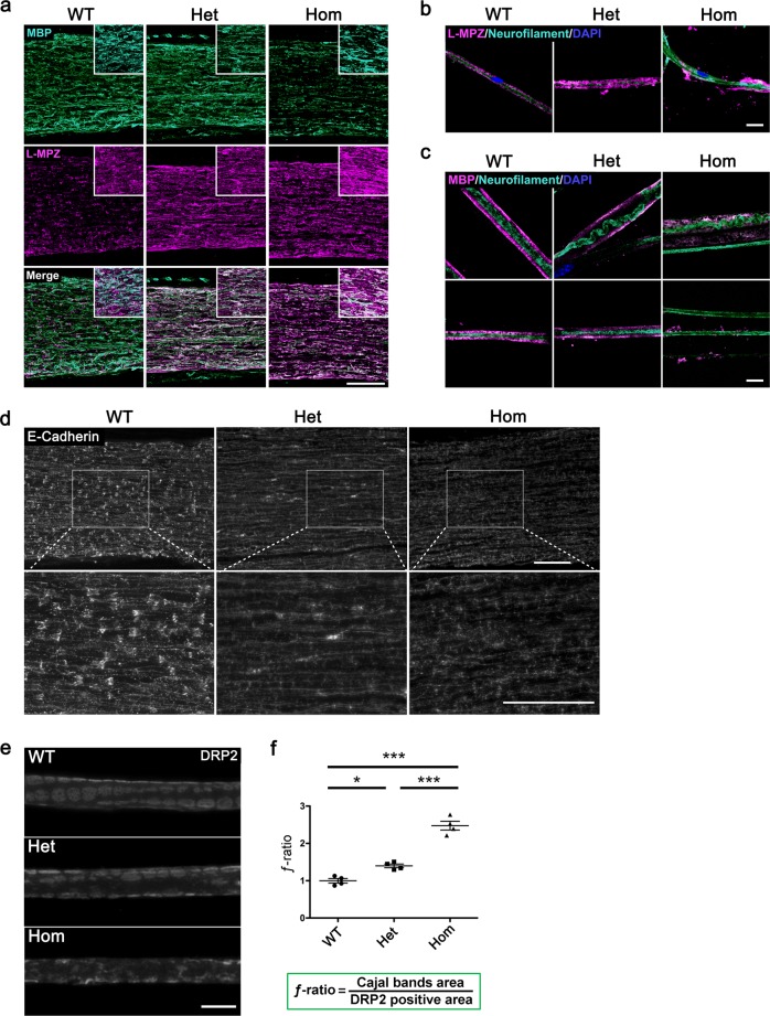
Fig. 2. Abnormal myelin structure in L-MPZ mice by immunohistological analyses.
a Double immunostaining patterns of sciatic nerve sections from 10-week-old L-MPZ wild-type (WT), heterozygote (Het), and homozygote (Hom) mice using anti-MBP (turquoise) and L-MPZ (magenta) antibodies. b, c Triple immunostaining patterns of teased sciatic nerve fibers in L-MPZ mice. Teased fibers were immunostained using anti-neurofilament (turquoise) and L-MPZ (magenta; b), or MBP (magenta; representative two images, c) with DAPI (blue). d Visualization of Schmidt–Lanterman incisures (SLIs). Immunostaining of sciatic nerve sections in L-MPZ mice was performed using anti-E-cadherin antibody (white). The white square is enlarged. e Cajal bands visualized as unstained areas by anti-DRP2 antibody staining (white). DRP2-positive flanking regions are shown as a cobblestone appearance. f For quantification, the ƒ-ratio (which indicates the ratio of Cajal band area [cytoplasm-containing channels] to DRP2 positive area [non-channel myelin surface]) was measured in teased sciatic nerve fibers. Bars, 100 µm (a, d) or 10 µm (b, c, e). *p < 0.05; ***p < 0.001 by one-way ANOVA with post-hoc Tukey’s test. Data are presented as mean ± SE of experiments. N in WT = 4, Het = 4, Hom = 4 (f). Data in f are presented as mean ± SE of four experiments.

